
I have always thought no connections existed between art and subjects like biology, mathematics and chemistry. I thought these subjects to be independent of one another. It was not until I read about the early experimentation of American artist Charles Csuri or the artwork of Joseph Scheer that I realized I had been creating art in classes like biology and chemistry. Charles Csuri used wave functions to digitally modify the reproduction of landscape, while Scheer used a scanner to scan the bodies of moths. When I first saw the artwork of Scheer it made me think of Lepidopterists, scientists that studies butterflies and moths, or even a collector.
But my favorite piece is Canogar hide 2 by Daniel Canogar. The portrait was created by inscribing many different fingerprints digitally; the prints blurred and overlapped creating only partial prints. Today fingering printing is one of the top methods in identifying someone. The fingerprint is so unique that you don’t share it with anyone else. However in Canogar’s piece he seems to have created a grand collage consisting of many different people; all the prints are blurred together, however, becoming an inseparable and unidentifiable piece of work. The work is the extreme contrast of what the fingerprint represents in our society today. Our fingerprint is our own individual identifier, but by making it unidentifiable, Canogar has made it anonymous. Scheer does the same thing in his pieces of work. The scientist or the collector usually took the moth itself or pictures of the moth and labeled it so that they could identify it; here Scheer reproduced the pictures but not the names, we are left with little other information but what we see on the page or in the picture. These artists have taken processes of identification and made them anonymous in their artwork.
I began to think of examples from my own life in which scientific processes could be made into art. One of the more prominent examples that comes to mind, is one of my biology labs. In this particular lab, we were looking at different types of cells.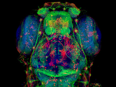 The cell had been stained with different florescent dyes. Each part of the cell could absorb a different dye because it’s density. We then used a confocal microscope connected to a computer to view the cell. The microscope would shine a light of different wave lengths, and which ever dye reacted to that wave length would reflect back a color and then we would photograph it with a computer program. This would be one repeatedly at different wavelengths to capture the different densities of the cell. After we were finished with this process, we would recreate the cell by overlapping the different photographs we had taken. The finished product created a beautiful 3D picture of the cell, with many colors over lapping, that looked it been created digitally on computer. Many scientists display their work at competitions like the Small World Microtography Competition. Scientist submit the pictures of their as forms of art1. It lead me to think that there may be many more scientific process that we are not exposed to that have the capacity to produce beautiful artwork.
Sources
Paul, Christiane. Digital Art. New York: Thames & Hudson, 2003.
1.http://scienceblogs.com/retrospectacle/2007/11/confocal_image_of_cochlea_wins.php